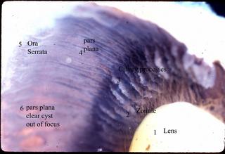
Click to enlarge the photo. The lens (1) is suspended by suspensory fibers to the ciliary body, the radial arrangement of suspensory fibers being termed the lens or ciliary zonule. These fibers are composed mainly of the protein fibrillin. Note that the pigment on the zonular fibers has allowed them to be visible as the span between both the anterior and posterior lens to the tips of the ciliary processes. The lens zonule holds the lens in place. Also note that some drop into the valleys between the ciliary processes to run their course to insert on the pars plana and some may even go to the retina (source of retinal zonular traction tufts). The ora serrata is where the retina joins the ciliary epithelium of the pars plana. The pars plana clear cyst is out of focus.
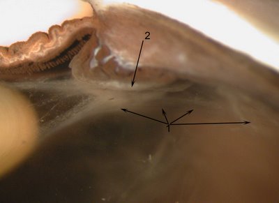
In the cross sectional photograph below, the zonular fibers can be seen from the origin on both the front and the back of the lens. The fibers from the lens connect to the ciliary body span fibers (arrow 2) like a suspension bridge and the extend all the way to the pars plana (arrow 1). Some of the fibers insert on the retina and are the origin of the zonular traction tufts that are a common finding in eyes. In this case material deposited on the lens makes the zonule visible. (<
NEXT Topic in Ocular Anatomy>
