LIGNEOUS CONJUNCTIVITISDefinition: Ligneous conjunctivitis is a woody induration of the eyelid accompanied by a plasminogen defect that leads to fibrin deposition and a recurrent pseudomembranous conjunctivitis.
Etiology:
Several mutations have been detected in the plasminogen gene of patients affected with ligneous conjunctivitis. The human plasminogen gene, located on chromosome 6, has a marked homology with the genes belonging to the plasminogen-apo(a) family, and with a number of pseudogenes and plasminogen-like genes located on chromosome 2. The result is that plasmin, the active form of enzyme that degrades fibrin, does not function and fibrin deposits accumulate.
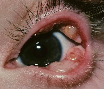
Clinical Presentation: The median age at diagnosis is about 5 years, and patients present with papillary white red lesions of firm consistency. Below is an image of lesions in both eyelids.
 Pathology:
 Fibrin is deposited in the substantia propria and epithelium may proliferate along the lobules of fibrin to become entrapped. The tissue may have a polypoid appearance and chronic inflammation and granulation tissue usually accompany the process. In the gross photograph islands of translucent granulation tissue and foci of inflammation (arrow 1) are surrounded by white fibrin (arrow 2).

Note the lobulated appearance of the mass.
Under the microscope the H&E stain shows the amorphous eosinophilic substance in the substantia propria.

The PTAH stain demonstrates the abundance of fibrin in the lesion (blue color).
 Treatment:
 The lesions generally respond well to fibrinolytic therapy, topical plasminogen and heparin.
